- Home
- / Services
- / Diagnostics
Diagnostics
1. Ultrasound (Sonography)
Ultrasound is a simple imaging technique which allows internal organs to be depicted on a monitor. The ultrasound head, which the physician places on the patient's body, sends sound waves into the body. These are reflected by the various organs, registered and translated into an image. This generates images of internal organs such as the pancreas or the liver, which can then be assessed by the physician.
The examination is painless and has no side effects; it can be repeated as often as necessary, but its informative value may be insufficient in certain cases, which is why other procedures are used.
2. Computer Tomography (CT)
Computer tomography works with X-rays. In questions of pancreatic disorders, its informative value is far superior to that of ultrasound.

As a rule, this procedure produces a very good image of the internal organs; the use of contrast media also allows determining the relationship of the tumour to certain other tissues, e.g. blood vessels or neighbouring organs. Prior to the examination, the patient has to drink a special liquid to increase contrast quality in the gastrointestinal tract. The actual examination is carried out in a tube-like structure through which the patient is passed automatically, lying on a scanning table. Some patients develop a hypersensitive reaction to the contrast medium which can manifest itself in a range of symptoms.
3. Magnetic Resonance Tomography (MRI)
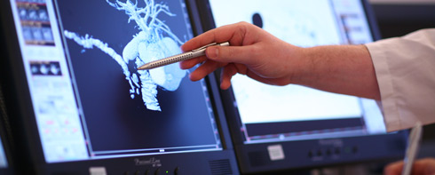
The MRI examination is similar to computer tomography and also carried out in a kind of tube. In this tube, sectional images of the body and the internal organs are taken. This procedure does not use X-rays, but works using changing magnetic fields. Individual departments, such as the University of Freiburg Department of Radiodiagnostics, are specialized in pancreatic imaging. Using specific techniques, they often successfully type a cystic tumour just from the image. In addition, an MRI scan can also produce a good image of the bile ducts and pancreatic duct (MRCP) and an exact depiction of vascular structures (MR Angiography).
4. Endoscopic Retrograde Cholangiopancreatography (ERCP)
In an ERCP procedure, a flexible telescope is passed through the mouth down to the duodenum (similar to a gastroscopy). This allows depiction of the mouths of the bile ducts and the pancreatic ducts into the intestines. Through a small catheter, a contrast medium is injected into the bile ducts and these thus indirectly depicted. The purpose of the ERCP is to gain a precise impression of the bile ducts and the pancreatic ducts. In some cases, the procedure is also used to ensure better drainage from the bile and pancreatic ducts into the intestines by inserting a small plastic tube (stent) into the bile duct.
Analysis of MRI

An MRI scan can produce a good image of the bile ducts and pancreatic duct (MRCP) and an exact depiction of vascular structures (MR Angiography).
